International Association of Professional Farriers


INTERNSHIP AT FORGING AHEAD - Report # 2 by Alan Baker RN BSN MBA
As an intern at Forging Ahead I spend a lot of time working with the veterinarians, vet techs, and equine nurses at the Marion DuPont Scott Equine Medical Center (EMC) in Leesburg, Va. They do amazing work helping acute/chronic laminitis, founder, and traumatic injuries. This article, along with several future articles, will focus on one of the many cases the EMC staff see on a daily basis. I must confess that as an apprentice farrier I have had tunnel vision during my assessment of the horses I've worked on prior to trimming and shoeing, focusing primarily on the horse's feet.When this particular assessment started at EMC, we were told that the horse wore size 5 hand forged shoes with studs for traction in the grass. I wasn't able to see any of the lameness or understand how the veterinarian was making his diagnosis of the different areas in question. The mare, a 15 year old draft-cross fox hunter, had a steady gait with no interference or observable foot issues. When I questioned the vet, he told me to stop watching the mare's feet and "look up". Lameness is not always a foot issue, but pinpointing the issue above the feet can be tough, as I'm about to explain.
Assessment: The veterinarian palpated all four lower legs, and then did a manual flexion/extension of the limbs, checking for tenderness and abnormalities. A walk/trot assessment in a straight line and in a circle going both directions was done. The mare was positive for both hock and upper limb flexions. There also appeared to be some right front leg discomfort. Observing the horse from both the front and the rear, it became clear that her right hip was distinctly higher than her left. This may be trauma or conformation related.
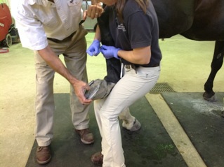
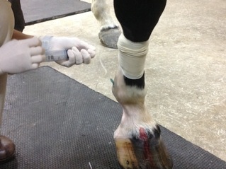
Procedure 1: After discussing treatment options with the owner, the veterinarian opted to begin his focus on the most suspicious cause of lameness by blocking the right hind leg suspensory ligament with Mepivacaine 2% (Carbocaine) to numb the nerve. The injection site was prepped by the vet tech using Hexadene (a topical antiseptic solution) prior to the procedure. Post procedure wait time was 10 minutes to determine effectiveness of the medication before reassessment.
Reassessment: There was no observable difference during the second walk/trot assessment. The mare continued to show grade 1/5 lameness post Mepivacaine injection. The right hind flexion was then repeated with no discernible improvement.
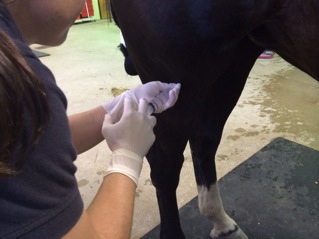
Procedure 2: After more discussion with the owner, the veterinarian moved up the leg to concentrate on the right stifle, blocking it with Mepivocaine 2% in three areas: the medial/lateral stifle (femoral tibial joint), and the anterior stifle (femoral patellar joint). Since this is a joint injection the vet tech prepped the injection site and surrounding area extensively with Hexadene to prevent infection in the joint which would cause serious problems and possibly wreck the horse permanently.
Reassessment: The mare was then walked around for approximately ten minutes r/t to her size in order to give the meds a chance to diffuse around the joint, but not so long that it could migrate to other areas which might cause an inaccurate walk/trot assessment. There was minimal to moderate change post injection of the stifle, except now there was increased lameness showing in her right front. Pain in the right front foot could have been masked by severe pain in the right stifle which had now slightly decreased. The veterinarian suspected the possibility of ringbone since this horse is a draft cross and decided radiographs needed to be taken.
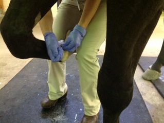
Procedure 3: The veterinarian blocked the abaxialsesamoid above the right front fetlock with an injection of Mepivacaine 2%. Prior to injection the site was prepped with Hexadine but not as vigorously as it would have been for a joint injection.
Reassessment: After waiting ten minutes the right hind was manually flexed by the veterinarian and the mare was put through her paces. Another round of walk/trot in a straight line and circles showed no visible change to either problem area.
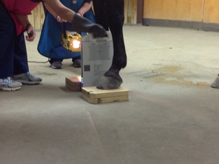
Procedure 4: The mare was sedated with xylazine. Radiographs revealed pinching of the distal medial pastern joint which sits down in the hoof capsule. Minor ring bone with observable roughness around the bones along with a visible cyst inside P2 was also revealed. (This is unusual and there's no specific treatment even if it's causing the pain as it is situated below the hoof capsule and would be very difficult to access.)
Procedure 5: Because of observable joint arthritis, the pastern and coffin joint were injected with Kenalog (steroid) and hyaluronic acid (a joint lubricating agent). The pastern bone cyst was treated with an IV infusion of Tildren (used for navicular disease and osteoarthritis and has an anti-inflammatory effect). Aseptic technique is very important during this procedure r/t joint injection. A tourniquet was applied on the right front lower leg at mid canon bone and Tildren was infused into the palmer digital vein.
This mare was unable to be reassessed R/T sedation. She was discharged after sedation wore off and she was able to safely travel. She will have a follow up appointment in two weeks to check for effectiveness of drug treatment.